At the University of Limerick, Clíona M. McCarthy, under the supervision of Dr. John J.E. Mulvihill, is leading the charge in pioneering the study of ex vivo tissues’ micromechanics.
© 2024 Optics11 Life B.V.
In the world of tissue mechanics, advanced technologies are constantly pushing the boundaries of research, which enable scientists to explore areas that were once out of reach. One such area is the micromechanics of fresh biological tissues, where Clíona M. McCarthy, under the supervision of Dr. John J.E. Mulvihill at the University of Limerick, is breaking new ground in her PhD thesis on ex vivo tissue1, such as colonic2 and vascular tissues, using the Chiaro indenter from Optics11 Life.
The shift from macroscale to microscale testing
During our conversation, McCarthy emphasized the limitations of traditional macroscale mechanical testing. “Macroscale testing often fails to detect regional mechanical variations in soft biological tissues,” she explained. Here is where microscale testing comes into play. “Microscale testing allows you to examine disease progression in healthy tissue, focusing on tiny changes in the tissue environment,” she added.
McCarthy also noted that while microscale testing tools were initially developed for engineered materials, the Chiaro indenter is specifically designed for soft, fresh biological tissues. “There’s definitely a shift towards microscale testing in both industry and academia across different types of materials – cells, tissues, hydrogels—and application areas,” she observed.
The importance of testing fresh tissues
McCarthy’s research relies heavily on fresh, unfixed tissues. Testing fixed or frozen tissues, such as carotid and femoral plaques, can introduce alterations that misrepresent in vivo physiological conditions. She added, “With Chiaro’s expanded stiffness range, I can quickly test several fresh plaques without processing them.”
This approach is critical because biological materials exhibit viscoelastic behavior. One of Chiaro’s standout features is the dynamic mechanical analysis (DMA) that assesses the viscoelastic properties of various ex vivo and engineered tissues to investigate disease progression, as modulus measurements differ between healthy and diseased tissues. McCarthy believes that DMA capability significantly enhances biomimetic models designed to mimic the mechanical properties of living tissues. “In the literature, most biomimetic modeling is based on purely elastic materials. I would expect better 3D biomimetic models with their viscoelastic properties evaluated,” she explained.
Streamlining the research process
Beyond its advanced testing capabilities, the Chiaro indenter also offers a user-friendly data visualization tool that McCarthy appreciates. “Most data processing is very manual and time-consuming, especially when calculating properties like Young’s modulus. Chiaro’s data viewer software simplifies this process, allowing me to focus on analyzing the data rather than spending hours processing it,” she shared.
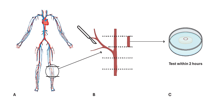
Note. Adapted from “Microindentation of fresh soft biological tissue: A rapid tissue sectioning and mounting protocol”, McCarthy, CM, 2024, PLoS One. 2024 Feb 29;19(2):e0297618. doi: 10.1371/journal.pone.0297618. © 2024 McCarthy et al. This is an open access article distributed under the terms of the CC BY 4.0.
Revolutionary micromechanical testing for disease development
As our conversation wrapped up, it was clear that Optics11 Life’s indenters are revolutionizing the mechanical testing of soft, fresh tissues at the microscale. Their dynamic analysis and efficient data visualization tools enhance accuracy and streamline research, making them invaluable assets for advancing preclinical studies on tissue mechanics during disease development.
But as McCarthy pointed out, it’s not just about technology. “Optics11 Life is committed to ensuring that users have an excellent understanding of what they are working with,” she said, praising the customer and application support provided by the qualified application specialists at Optics11 Life.
References
1 McCarthy CM, et al. Comparison of macroscale and microscale mechanical properties of fresh and fixed-frozen porcine colonic tissue. J Mech Behav Biomed Mater. 2023 Feb;138:105599. doi: 10.1016/j.jmbbm.2022.105599.
2 McCarthy CM, et al. Microindentation of fresh soft biological tissue: A rapid tissue sectioning and mounting protocol. PLoS One. 2024 Feb 29;19(2):e0297618. doi: 10.1371/journal.pone.0297618.